Female Reproductive Anatomy
The primary purpose of our reproductive system is to pass on our genes (and those of our partners) by conceiving and birthing a child. The reproductive system is made up of external and internal anatomy.
External

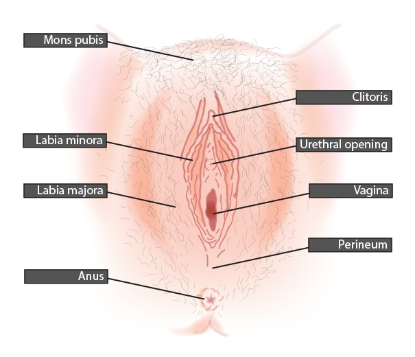
Our external anatomy is known as the VULVA. It includes
MONS PUBIS: Mound of fatty tissue above the pubic bone.
CLITORIS: A ball of over 8,000 sensitive nerve endings – designed purely to provide pleasure. The clitoris is a much larger structure than we can see from the surface. Internally, the clitoris extends through and behind the labia – it is comprised of erectile tissue which swells with blood when we are aroused.
CLITORAL HOOD: A protective hood of skin that houses the clitoris.
URETHRAL OPENING: This is where urine is expelled after travelling from the bladder through the urethra.
VAGINAL OPENING: The vaginal opening is where the vaginal canal exits the body.
BARTHOLINS GLANDS: On each side of the base of the vagina are the Bartholins glands – two pea-sized glands that secrete lubricative fluid when we are sexually aroused. Allows for easier penetration by the penis.
PERINEUM: Area of smooth skin that connects the vulva to the anus.
LABIA MINORA: Inner vaginal lips. These vary widely between women. It is normal for the labia minora to extend past the lips of the labia majora, to be asymmetrical, to be of differing lengths, to be a different colour than the rest of the vulva, and even to be barely visible in some cases. All variations are normal.
LABIA MAJORA: The fatty tissue that encompasses the smaller labia minora.
Internal

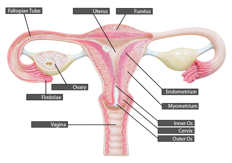
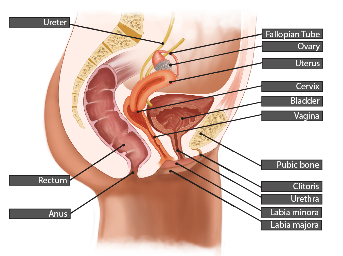
VAGINA: The vagina is a muscular tube that connects our uterus to our vaginal opening. It can expand to accommodate a mans penis and even a baby during childbirth. It allows menstrual blood, cervical fluid and other vaginal discharge to exit the body.
CERVIX: The cervix is the lower 1/3 of the uterus and is approximately 3cm long. The internal os (internal opening) opens into the uterus, and the external os (opening) opens into the vagina. Ovarian hormones cause the cervix to lower or retreat higher into the vagina and change in openness and firmness at different points of the menstrual cycle. Ovarian hormones also cause the cervix to secrete different types of cervical fluid at different times of the menstrual cycle. These secretions are produced from crypts that line the internal passage of the cervix.
UTERUS: Shaped like an upside-down pear and situated between the bladder and the rectum. The uterus has an internal lining called the endometrium. Each menstrual cycle, the endometrium grows and thickens in response to ovarian hormones. If a pregnancy does not occur, the endometrium sloughs away as menstrual bleeding. In most women, the uterus is tilted slightly foward over the bladder. Around 20% of women have a retroverted uterus that tilts backward instead.
FALLOPIAN TUBES: The fallopian tubes extend out from each upper corner of the uterus toward the ovaries. They are mobile structures around 10cm long. They culminate in the FIMBRIAE which are groups of 10-15 finger-like projects that assist with collecting an egg from the ovary at ovulation. The fallopian tubes are lined with tiny hair-like projections called cilia. The cilia beat in a wavelike motion to help transport a fertilised egg toward the uterus where it can implant into the endometrium.
OVARIES: The ovaries are each connected to the uterus via the ovarian ligament. The ovaries are not connected to the fallopian tubes; however, the fimbriae hover very closely around the ovaries. The ovaries are a whitish colour and each around 3.5cm long (the size of an unshelled almond). The ovaries are the site of the development and release of an egg at ovulation, and the ovarian hormones oestrogen and progesterone.

